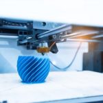
The 3D printing technology makes the dentistry industry a yet more effective & convenient service by bringing in rapid advances in technology. The impact of 3D printing or additive manufacturing in digital dentistry is quite important for providing customized solutions for dental problems & issues. The 3D printing market in the dentistry sector was valued at $1.8 billion in 2020 and is estimated to reach $6.5 billion by 2025, at a CAGR of 28.8%.
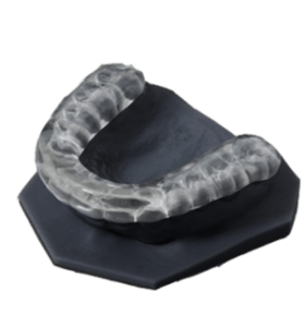
The role of the 3D printing market in digital dentistry is a massive game-changer because of its rapid high performance in faster production, increased efficiency, and improved quality. Dental clinics and labs all around the world use 3D printers for producing products such as Surgical crowns and bridges, Dentures, Retainers, Splints, Clear Aligner molds, Orthodontic models, Surgical guides, sports guards, etc.,
What is Digital Dentistry?
Digital dentistry is a dental technology incorporated with computer software components that provide validated end-to-end workflow. And, this digital workflow results in higher-end and complex product production at a low cost and short duration; whereas, traditional dental mediums use mechanical or electrical procedures for manufacturing dental products which consume more time and labor work.
As a result, many dental professionals joined hands with the digital dentistry evolution to provide upgraded solutions to patients for traditional dental problems. The following digital technologies of the dental industry areas are listed below.
- Computer-aided design or computer-aided manufacturing (CAD / CAM).
- Computer-aided implant dentistry.
- Digital Radiography.
- Cone-beamed computed tomography imaging (CBCT).
- Lasers.
Computer-aided design or computer-aided manufacturing (CAD / CAM)
Computer-aided design or computer-aided manufacturing (CAD/CAM) technology has played a crucial role in digital dentistry over the past 25 years. This technological development in the dentistry sector provides us with better working conditions and increased comfort for both dentists and patients.
The dental laboratories started fabricating restorations, even full mouth restorations, and complete dentures. Computer-aided manufacturing (CAM) uses generated paths to shape a restoration. It uses specially designated software called CAD software provided by the manufacturers for designing countless 3D dental restorations on computers.
The first process is data acquisition, which involves digitizing clinical data. Followed by this process, jaw and tooth structures of the mouth are measured by the intraoral camera, or by scanning plastic impressions. CAD technology creates virtual objects using graphic representation and finally virtual model appears on the screen after digitization.
Computer-aided manufacturing (CAM) transforms the virtual object into a material object. The final process is the Numerical control machine tools, which are responsible for manufacturing, using programmable machine tools fitted with numerical control. This manufacturing method is either done by subtraction method (by removing materials) or additive method (by adding materials).
And, this technology helps to overcome the several drawbacks associated with wax models. The drawbacks that are to be rectified include eliminating shrinkage and expansions, avoiding shrinkage and porosities associated with the casting procedure, and making restoration of tooth easier, and faster with accurate fit.
A study in 2018 remarked that more than 30,000 dentists around the world own scanning and milling machines. And, also this study predicted that more than 15 million restorations have been completed worldwide.
With the help of CAD/CAM technologies, dental professionals can mostly provide restorations to patients within the same day. And, CAD-CAM is also used in orthodontics through Invisalign retainers.
Computer-aided implant dentistry
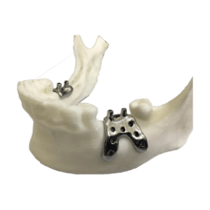
Performing implant prosthetic surgery has never been easier. Implant 3D is the software that enables the user to simulate the position of implants on two-dimensional and three-dimensional models.
Some of the advanced implant technologies are Three-dimensional computed tomography (CT) imaging, Computer-guided implant surgery (CGIS), CT-based implant-planning software, Computer-aided-design/computer-aided-manufacturing (CAD/CAM) technology, Computer navigated implant surgery (CNIS), and Robotic-Implant-dentistry.
The Computed Tomography-based implant-planning software allows an undistorted three-dimensional visualization of the jawbone in axial, sagittal, coronal, panoramic, and cross-sectional views. Even it produces three-dimensional reformatted reconstructions. The main advantages of implant-planning software are,
- Digital planning
- Allows the predetermination of the size of the implant
- Complications in the selection of implant size can be easily rectified
- Provisional restoration
- Assists in anticipation, guiding, and planning of procedures like alveolectomy.
Computer-Guided Implant Surgery (CGIS) is a static system that makes use of a static surgical template or guides to produce the virtual implant position. Based on CAD/CAM technology, it can be categorized into two types. The advantages of Computer-guided implant surgery include,
- Helping the surgeon to place the implant in the exact location based on a virtual treatment plan
- Allows flapless surgery
- Aids in the preservation of hard and soft tissue
- Precisely guides the osteotomy drills.
A few drawbacks associated with CGIS include fracture of the surgical guide and secondary thermal injury.
Computer Navigated Implant Surgery (CNIS) is a dynamic system that reproduces virtual implant position directly from CT data with the optical bur tracking system which involves the use of a surgical navigation system. It has many advantages over CGIS. The major advantage is that it allows intraoperative changes that are to be done in the implant position so the virtual surgical plan can be altered or modified during surgery. The CNIS overcomes the limitations of CGIS like secondary thermal injury, displacement, fracture of guide, etc.
Digital Radiography
The usage of digital dental radiography has increased in recent years to better detect, diagnose, treat, and monitor oral conditions and diseases. In digital radiography, traditional photographic X-ray film is replaced with digital X-ray sensors, which results in enhanced computer images of teeth, gums, and other oral structures and conditions.
Digital dental images in digital radiography can be acquired through three methods. The following methods are the direct method, indirect method, and semi-indirect method. In the direct method, an electronic sensor is placed in the mouth to record the images of it. The X-ray film scanner is used in the indirect method to view the traditional dental X-rays as digital images. The semi-indirect digital technique combines both sensor and scanner to convert traditional dental X-rays into a digital film.
Types of digital dental radiographs
Digital dental radiographs are categorized into Intraoral (Inside) X-rays and Extraoral (Outside) X-rays. Intraoral X-rays provide great details and are used to detect cavities, check and monitor the developing teeth and bone health. Whereas, Extraoral X-rays do not provide details like intraoral X-rays but they are used to detect impacted teeth, monitor jaw growth, and jaw development. It is mainly used to identify potential problems between teeth, jaws, temporomandibular joints (TMJ), or other facial bones.
Intraoral X-rays are categorized into two types, which are Bitewing X-rays and Periapical (limited) X-rays. In the Bitewing X-rays, the images show details of the upper and lower teeth in one area of the mouth. Each bitewing shows a tooth from its crown (top) to about the level of the supporting bone and these bitewing x-rays are used to determine the fit of dental crowns as well as the marginal integrity of tooth fillings. In the Periapical (limited) X-rays, the whole tooth can be viewed from the crown to beyond the root tips to the supporting bone. It is used to detect root structures and surrounding bone structure abnormalities.
The types of Extraoral X-rays include Panoramic (Panorex) X-rays, Multi-slice computed tomography (MCT), Cephalometric projections, and Sialography. Panoramic X-rays require a machine to show the entire mouth including all the teeth in the upper and lower jaw. These are used to plan treatment for dental implants, detect impacted wisdom teeth and jaw problems.
The major use of Panoramic X-rays is to identify unrecognizable bodies after fires, crashes in the forensic and legal departments. Multi-Slice computed tomography is used to view a particular layer of the mouth, this type is mainly helpful in examining complex structures. Cephalometric projections tend to show the entire head, help in examining the teeth with a patient’s jaw and profile. In Sialography, dye (radiopaque contrast agent) is injected into the salivary gland to visualize them on the X-ray film. It is used to identify salivary gland problems, such as blockages or Sjogren’s syndrome.
Cone-beam computed tomography imaging (CBCT)
Cone-beam systems are used to capture data from the patient using a cone-shaped X-ray beam. The three-dimensional (3D) images of the following regions of the patient’s anatomy are reconstructed from the above data. Cone-beam computed tomography imaging is a fast, non-invasive way of answering several clinical questions.
Cone-beam computed tomography images are dimensionally accurate and cannot be controlled by anatomic noise. It is two times better than periapical radiography in detecting periapical radiolucencies. Due to the improved sensitivity, it is considered to be a diagnostic tool.
As a diagnostic tool, Cone-beam computed tomography imaging (CBCT) solves the problems by reducing the difficulty level of localizing symptoms, areas subject to anatomic noise, the workup of traumatic dental injuries, and the assessment of resorptive lesions.
Some of the benefits of Cone-beamed systems include,
- A focused X-ray beam reduces scatter radiation resulting in better image quality
- A single scan makes a wide variety of views and angles to provide a more complete evaluation
- Ability to image bone and soft tissues at the same time.
Lasers
The term LASER is an acronym for “Light Amplification by the Stimulated Emission of Radiation” In digital dentistry, a laser is an innovative tool in modern dental practice to create the first 3D layer of a solid object. Laser light is a monochromatic light with three principal parts and a single wavelength of light. The three principal parts of laser light are an energy source, an active lasing medium, and two or more mirrors from an optical cavity or resonator.
In the dental laser, amplification occurs by supplying energy to the laser system via a pumping mechanism, and this energy is pumped into an active medium contained within an optical resonator, which led to producing spontaneous emission of photons. The dental laser lights are delivered from the laser to the target tissue through a fiberoptic cable or articulated arm.
The uses of laser in hard tissue application are caries prevention, bleaching, cavity preparation, dentinal hypersensitivity, growth modulation, restorative removal, and diagnostic purposes. In soft tissue application, the laser is used for wound healing, removal of hyperplastic tissue to uncover the impacted tooth, photodynamic therapy for malignancies, and photostimulation of the herpetic lesion.
Laser in dental practice can be classified into various methods, depending upon the lasing medium used, such as gas laser, and solid laser; and based on tissue applicability, hard tissue, and soft tissue lasers. Some of the lasers in dental practice used by dental professionals include Carbon Dioxide Laser, Neodymium Yttrium Aluminium Garnet Laser, Erbium Laser, and Diode Laser.
Benefits of 3D Printing in Dentistry
What exactly are the benefits or advantages that 3D printing provides when it comes to dentistry?
- Digital dentistry helps dentists to get a clear view of the image which leads to improved diagnostic ability and faster treatment. This 3D technology and optimized solutions allow people to get the best possible care for their teeth and gums.
- The traditional tray-and-putty dental impressions are being replaced by digital impressions, which results in faster turnaround times, real-time prep feedback, minimized remarks, and smoother workflow.
- Digital scanners (i.e) an Intraoral scanner offer a much more efficient and convenient dental experience than the conventional molded impression, eliminate the number of steps in making restoration, and also minimize the risk of errors.
- With digital impressions, dental professionals no longer feel the need to coax patients into taking an impression. The digital impression has drastically improved the patient experience and helped in improving patient relationships and patient care.
- CAD/CAM restoration is preferred because it creates a natural appearance, and it improves the quality of restoration. It also produces dental restorations in a variety of shades, sizes, and colors. The advancements in technology can be used in cosmetic dentistry procedures such as onlays, inlays, dentures, and veneers.
- Other benefits of digital dentistry include the elimination of material inaccuracies for fewer restoration flaws, it can be stored electronically which supports a paper-free environment or green dentistry, less chair time, and no need for distasteful impressions saves time and money.
Digital dentistry is growing at a fast pace and continues to step forward and make the delivery of dental care more comfortable, predictable, and precise. The size of the U.S. digital dentistry market is expected to reach $986 million in 2021, with the CAD/CAM material market expected to experience faster growth.
Due to the COVID-19 impact, the size of the U.S. dentistry market is predicted to reach $ 1.37 billion over the forecast period in 2027. The size of the European digital dentistry market is expected to reach €578.7 million in 2021, with the CAD/CAM material market expected to experience the highest growth. The size of the European dentistry market is predicted to reach €1.4 billion over the forecast period in 2027 due to the COVID-19 impact.